Who understands X-rays? What is radiography? In what cases is the procedure performed?
Radiography is a specific type of examination internal systems and organs of the human body. When it is carried out, a projection of the area under study is created on film or special paper. This is facilitated by X-rays. Based on such a projection, a specialist can draw certain conclusions.
Radiography is the first medical imaging technique. It allows you to obtain images of organs and tissues for their study during the patient’s lifetime.
Radiography is a diagnostic method that was discovered by the German physicist Wilhelm Conrad Roentgen in 1895. He recorded the ability of x-ray radiation to darken a photographic plate.
Description of the diagnostic method
What is radiography based on? This study is made possible thanks to the high penetrating power of X-rays, which are created by a sensor of a special device.
Such radiation passes through the tissues of the human body. At the same time, it not only ionizes cells, but also lingers in them. The volume of such presence of X-rays in tissues varies. This allows a black and white image of the area under study to appear on film. Bone tissue is more radiopaque. That is why in the photographs her image appears in light colors. Dark areas of the film represent soft tissue. These areas absorb X-rays very poorly.

It is clear that radiography is the study of three-dimensional objects. However, on film, all images come out flat. In this regard, photographs are taken in at least 2 projections. This allows you to accurately detect the location of the source of pathology.
Advantages of the technique
What are positive sides, which radiography of organs has? They are as follows:
Ease of conducting research;
- wide availability of the method;
- no need (in most cases) for special preparation of patients;
- relatively low cost (except for studies whose results are obtained digitally);
- absence of operator-dependence, which facilitates the consideration of the data obtained by specialists during consultations.
Negative aspects of the technique
Despite the fact that radiographic examinations are widespread in modern medicine, they still have some disadvantages:
The resulting image is “frozen”, which greatly complicates the diagnosis of functioning internal organs;
- X-rays have a harmful ionizing effect on the human body;
- the results obtained have low information content when compared with the latest tomographic methods;
- when examining soft tissues, there is a need to use special contrast agents.
Prevalence of the method
Thanks to the discovery of X-ray radiation, medicine has made a significant breakthrough in the field of diagnostics. huge amount diseases that, before the discovery of the German physicist, were detected only at a late stage, which made it difficult or impossible to treat the disease.
Today, X-rays can be taken in most clinics and hospitals where special equipment is available. With the help of the research carried out in the most short time the diagnosis is clarified and necessary plan treatment.

In addition, the doctor sends his patients for x-rays so that they undergo a preventive examination. Sometimes this helps to diagnose serious pathologies at the earliest stages of their development. The most famous and widespread type of such research is fluorography. The purpose of its implementation lies in the possibility of early diagnosis of pulmonary tuberculosis.
Classification
There are various x-ray examination techniques, which differ in the way they record the resulting image. So, they distinguish:
1. Classic radiography. It allows you to obtain an image using direct impact of ionizing rays on the film.
2. Fluorography. When using this type of technique, the image appears on the monitor screen, from which it is printed on small-format film.
3. Digital X-ray. The result of this study is black and white image. The picture is on digital media.
4. Electroradiography. During this study, the image is captured on special plates and then transferred to paper.
5. Teleradiography. This study uses a special television system that displays images on a television screen.
6. X-ray. With this technique, the desired area can be viewed on a fluorescent screen.
Digital radiography most accurately reflects the picture of the study area. This technique greatly facilitates the diagnosis. And this allows you to more accurately select a treatment regimen.
Object of research
Depending on which organ or system is being diagnosed, the following research options are distinguished:
X-ray of the spinal column and limbs;
- chest;
- teeth (intraoral, extraoral, orthopantomography);
- breast (mammography);
- colon (irrigoscopy);
- duodenum and stomach (gastroduodenography);
- gallbladder and biliary tract (cholecystography and choleography);
- uterus (metrosalpinography).
Indications
The doctor refers his patients to x-rays, as well as to other x-ray examinations. He does this only if there is evidence, of which there is a great variety. The main ones:
Carrying out diagnostics of pathologies of internal organs and skeleton;
- checking the effectiveness of the treatment and determining its negative consequences;
- monitoring of installed tubes and catheters.
Contraindications
Before sending a patient for an x-ray, the doctor must find out whether the patient has serious reasons not to undergo this study. But it cannot be carried out in the following pathologies and conditions:
Active forms of tuberculosis;
- disorders of the thyroid gland;
- general serious condition of the patient;
- pregnancy (for women expecting a child, radiography is performed only if there are vital indications);
- breastfeeding (in cases where it is necessary to administer a contrast agent);
- renal and heart failure (contraindication also applies to contrast);
- bleeding;
- allergies to substances containing iodine (if it is necessary to introduce contrast elements).
Decoding the results
How to correctly read the resulting radiographic projections? This can only be done by a specialist with the necessary qualifications. Such work cannot be performed by a person ignorant in this area.
Those images that are the result of radiography are negatives with light areas of denser structures of the body and dark ones, which indicates the presence of soft tissue in this place. Deciphering each area of the body is done according to certain rules. So, when examining a chest X-ray, a specialist should evaluate the relative position, as well as the structural features of the heart, lungs and mediastinum. In addition, the collarbones and ribs are examined to identify cracks and fractures. All obtained parameters are assessed based on the patient’s age.

In order to make a final diagnosis, a doctor, as a rule, does not have enough of one image. In addition to radiography, the presence of pathology can be determined based on examination data, interviews, as well as the results of various instrumental and laboratory examination methods.
X-ray of the spine
Often the doctor sends his patient for examination of this part of the body in case of injury and to make the necessary diagnosis. X-ray of the spine is considered the most conservative method. To carry it out, no preliminary preparation is required from a person.
X-ray of the spine can give an objective picture only if it is performed in two projections. The first image should be taken with the patient lying on his back. The second one is lateral. This is a photo of the lumbosacral region.
An X-ray of the spine is performed if pain occurs in the back. In case of emergency, such a procedure is carried out at home.
The reason for examining the cervical spine is severe headaches, as well as dizziness with rapid turns of the neck. Such fluoroscopy is performed in two projections. Often, in order to obtain more detailed information, images are taken through the patient's open mouth.
Indications for performing x-rays of the thoracic spine are pain in the chest that occurs when bending or turning. Distinctive feature Such a study consists of taking a picture in three projections: side, back and front.
In order for a survey radiography of the coccyx and lumbosacral region to be performed, preparatory measures will be required. First of all, this is a diet that must be followed for several days (usually two) preceding the examination. It consists of eliminating from the daily diet those foods that cause gas formation in the intestines. In this case, the patient should not eat cabbage and potatoes, consume rye bread, milk and beans.

The studies themselves are performed only on an empty stomach and with cleansed intestines. If the patient is not properly prepared, accumulations of intestinal gases that do not allow X-rays to pass through can give a unclear picture of the area being examined.
The result of the x-ray will be an image in which the specialist will be able to see the person’s spinal pathologies. These are osteochondrosis and vertebral hernia, spinal tuberculosis, its curvature, etc.
Joint studies
Often, a doctor needs to make a diagnosis for existing disorders of the osteoarticular system. For this, the patient is prescribed radiography of the joints. Only in the images obtained during such a study can one see the following signs of pathology:
Calcium deposit areas;
- bone growths occurring on the edge of the cartilage;
- violation of the conformity of joint surfaces.
X-rays help the doctor identify existing problems to make an accurate diagnosis, as well as determine the type of treatment and plan it.
The doctor may order x-rays:
Ankle joint;
- knee joint;
- hip joint;
- elbow joint;
- shoulder joint;
- temporomandibular joint.
X-ray of the stomach
This research method allows us to identify numerous diseases of this important digestive organ, as well as the presence of its functional disorders.

X-ray of the stomach helps determine:
Peptic ulcer;
- malignant and benign neoplasms;
- diverticula (protrusion of the wall of this organ in the form of a bag).
X-ray of the stomach helps determine its size and position, the integrity of the wall and many other parameters. In order to examine this hollow organ, a contrast procedure is required. Barium salts suspended in water are used as a substance that does not transmit x-rays. Sometimes gas serves as a contrast.
Lung studies
This diagnostic method, in addition to general indications, is applied to a certain category of the population. These are, for example, people who constantly experience hazardous production conditions: masons and miners, enterprise workers chemical industry etc.

X-ray of the lungs reveals:
Pneumonia of the lungs;
- hydrotax (accumulation of fluid in the pulmonary tract due to liver cirrhosis, ascites, heart failure);
- pneumothorax (mechanical damage to lung tissue);
- chronic diseases (atypical pneumonia, silicosis, tuberculosis, lupus erythematosus, etc.).
Only radiography will allow timely recognition of the onset of the above pathologies and selection required course treatment.
To believe that THIS could be inside a living person, you need to see their X-rays with your own eyes. We bring to your attention the strangest X-ray images in the history of medicine, from which you feel creepy and scared, and sometimes you experience surprise or even real shock.
(Total 20 photos)
1. The skull of a Chinese man who was shot in the head with a pneumatic hammer.

2. The stomach of a patient who swallowed two forks, a ballpoint pen and a toothbrush.

3. Antique x-ray of the foot of a Boer War soldier (1899-1902) with a gunshot wound. The bullet lodged in the metatarsal bone between the big and second toes.
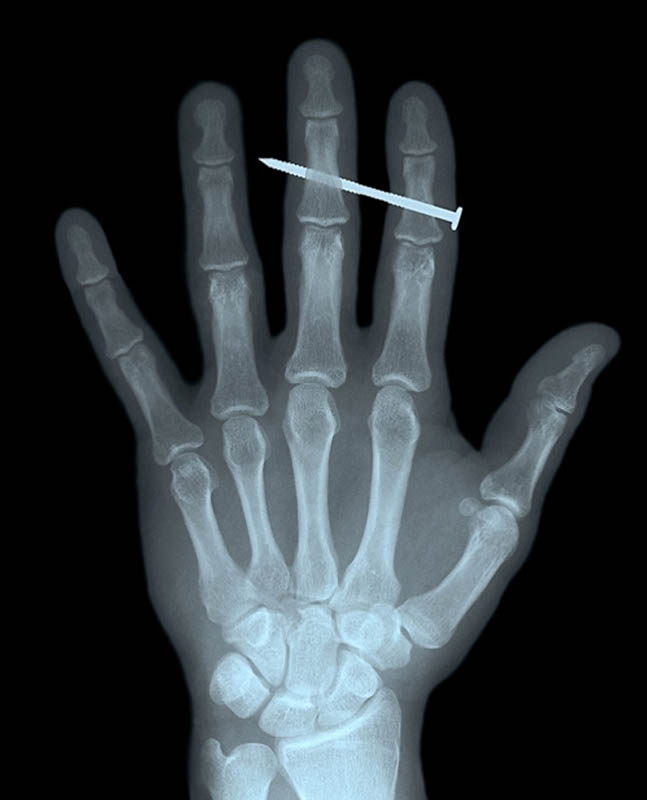
4. Nail in the bones of the index and middle fingers of an adult man.

5. Color photograph of objects that the patient swallowed and which became lodged in his intestines, including a spoon and blade.

6. A pin in a woman's throat.

7. X-ray of a patient who stepped on a fork.

8. Color x-ray of the stomach of a patient who swallowed a razor (center left) and blades (top right).
9. A torn finger of a patient who fought with a man armed with a knife.

10. Another patient who stepped on a fork.

11. A harpoon spear hit a 16-year-old boy in the head while fishing.
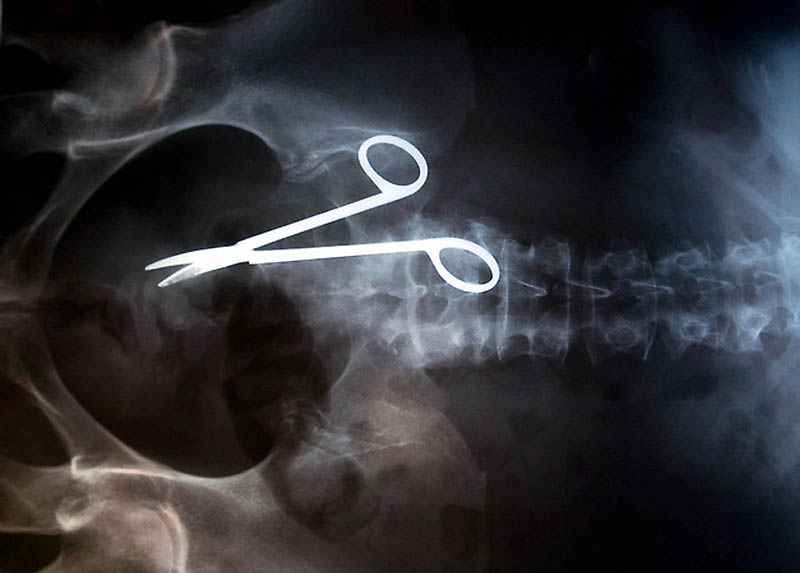
12. Surgical scissors, accidentally forgotten in the patient’s body after surgery. The scissors were discovered only 18 months after the operation, because the woman complained of constant pain in the intestines.

13. Nail in a human skull - a patient accidentally shot himself with a pneumatic hammer. He didn’t even realize that he had shot himself - the 10-meter nail was discovered only 6 days later.

14. Mobile phone in the intestine of a prisoner.

15. Knife in the head of a 10-year-old boy. The boy survived.
X-ray of the lungs is a summation image of the soft tissues of the chest. Along the path of X-rays, some structures absorb and others reflect radiation. Such a game is displayed on x-ray film or digital media.
A radiologist reads an x-ray consisting of a complex of white and gray shadows. Their combination forms an image, which a specialist deciphers and makes a description.
Our specialists are ready to interpret readers’ X-ray images for free. We also suggest that you carefully understand on your own the complex of X-ray darkening and clearing.
X-rays of the lungs are normal
X-ray images of the lungs (chest organs) are analyzed according to the “PoChiFora and InRiCoS” scheme. How to decipher these terms:
- Po – position;
- Chi – number;
- Fo – form;
- Ra – dimensions;
- In – intensity;
- Ri – drawing;
- Co-circuits;
- C – displacement.
This algorithm is taught to medical university students preparing to become radiologists.
Consider, for example, an x-ray of a normal lung:
It visualizes a lot of darkening and lightening (white and black), which can intimidate readers. In fact, this radiograph is easy to decipher (see next image)

All anatomical structures are labeled on the radiograph to make it easy for readers to understand. We suggest you remember the intensity of the lung fields. The norm does not imply the presence of pathological darkening (white) and lightening (dark), which are not in the image.
If you get your eye on it, you will learn to clearly distinguish normal from pathology.
X-ray of healthy lungs, how to read
X-rays of healthy lungs should be described according to the classical standard. First, entries are made about pathological X-ray syndromes, then the pulmonary fields, roots, domes of the diaphragm, costophrenic sinuses, cardiac shadow and soft tissues.
Keeping fit is part of the daily life of a modern person. Running or Pilates, karate or strength training - everyone chooses the type of activity that suits them. Unfortunately, sports are sometimes unsafe and injuries occur, but any coach can confirm that you should not be afraid of them. Thanks to modern diagnostic methods, it is possible to detect almost any “problem” in the human body and begin timely treatment. One of the most effective ways diagnostics is radiology. Based on the analysis of the X-ray image, the doctor will quickly and with a high degree of accuracy detect the problem.
X-ray: what does it show and what does it look like?
More than a hundred years have passed since the discovery of X-rays, but X-ray diagnostics still remains not only convenient and relevant, but sometimes the only possible method of diagnosis. Thanks to this study bone fractures can be diagnosed (X-rays for fractures are taken in frontal and lateral projections). The x-ray also clearly shows the pathology of the joints: arthritis, arthrosis, dislocations. In order to diagnose tuberculosis, fluorography is sometimes sufficient, but if the doctor has doubts when reading the image, he may prescribe an additional x-ray examination. X-rays are also used to diagnose diseases such as pneumonia, intestinal obstruction (the intestines are examined with contrast, the patient has to drink a barium sulfate suspension), neoplasms (both malignant and benign), aneurysms, spinal pathologies and some heart diseases. Also, thanks to this study, it is possible to determine the presence of a foreign body in respiratory tract or stomach.
What is an x-ray? Probably each of us has seen it at least once in our lives - it is a black and white image of the internal structures of the body, reminiscent of an ordinary negative. The light areas of the image are characteristic of the denser parts of our body, and the dark areas are characteristic of soft organs and hollow structures, such as the lungs. Based on the nature of the brightening and darkening, the doctor makes a diagnosis.
Previously, images were projected only onto a special light-sensitive film, but with the development of digital radiography, it became possible to obtain images in digital format. That is why in Lately, this primarily concerns private clinics; the patient increasingly receives not a film image, but a disk or flash card with the results of the study.
How is the fluoroscopy procedure performed?
X-rays are not only painless, but also, contrary to popular belief, a safe procedure. The dose of radiation that a person receives during fluoroscopy is very small and completely harmless.
As a rule, no preparation is required for an x-ray - you just need to follow the doctor's instructions: wear a protective apron that covers your reproductive organs and do not move while the x-ray machine takes the picture. However, in some cases, preparation is still needed: for example, when the patient needs to have an x-ray of the chest, spine or gastrointestinal tract. In order for the images to be as clear as possible, three days before the examination date the person will be asked to follow a special diet: exclude from the diet foods such as milk, brown bread, fresh cabbage, potatoes, beans and other foods that can cause flatulence. X-rays of the spine are performed only on an empty stomach, and the last meal can be no later than seven o'clock in the evening the day before the procedure.
How is an x-ray taken?
During the study, ionizing radiation passes through the human body. Soft tissues transmit rays, while dense tissues block them. The rays passing through the patient's body are recorded by a detector. When using analog devices, the detector is a fluorescent screen or film onto which the image is directly projected. The screen can also play the role of a kind of amplifier of received signals. After converting the radiation into an image using a special optical system, the latter can be recorded by a television camera and shown on a monitor (indirect analogue method). In the case of digital equipment, the data is recorded by the receiver and immediately converted into binary code, displayed on the computer screen. A digital photograph can be recorded on magnetic media, disk, or the image can be displayed on film.
As a result of all these manipulations, a planar black and white image of anatomical structures is obtained. Based on the shadows and light areas in the image, the doctor “reads” it and then draws a conclusion about the condition of certain internal organs.
The most modern and safest method today is digital fluorography - during its implementation the patient receives a radiation dose one hundred times less than during radiography. The radiation dose will be only 0.015 mSv, with a preventive dose rate of 1 mSv. However, the resolution of such a fluorograph is still inferior to digital radiography: on an X-ray of the lungs, the doctor will be able to see shadows measuring 2 mm, while a fluorographic study will show only shadows of at least 5 mm.
How to take an x-ray correctly and what determines the clarity of the image?
The clarity of an x-ray depends on several factors. These include the equipment on which the procedure is carried out and the correctness of the examination itself. So, for example, if the patient does not move while the image is being taken, the contours of the internal organs will be blurred and the doctor will not be able to clearly read the image.
If the doctor considers that one image is not enough to make an accurate diagnosis, he can prescribe additional x-ray examinations to the patient: take a photo of the desired organ in several projections: postero-anterior, anteroposterior, lateral or targeted.
For example, during a posteroanterior projection of the thoracic region or spine, the patient stands, his chin is fixed, and his breathing is held during the image. The anterior-posterior projection is done in the supine position and with a deep breath.
Lateral projection is often prescribed by a doctor if lung disease is suspected. It is done as follows: the patient is asked to lie down with his hands behind his head. His left or right side is fixed, breathing is held, and then a deep breath is taken. Also, the lateral projection is often used in determining sports injuries: for example, sprains, joint damage. During the procedure, the person will need to bear weight on the affected leg.
This is interesting
At the beginning of the 20th century, a new trend arose: the fashion for x-rays. Every self-respecting fashionista simply had to have a photo of his own bones at home - arms, legs, skull. In large cities, so-called studios were opened en masse, where everyone could take a photo of any part of their body. Since the dangers of X-rays were unknown at that time, even pregnant women came to the studio to “photograph” their unborn child. The pictures were expensive, and those who did not have enough money were given the opportunity to simply “shine” in front of the screen - by the way, this is how the world learned about the deformations of the ribs caused by wearing a corset.
X-ray image evaluation
When interpreting an X-ray image, the doctor takes into account the fact that it is formed by a diverging beam of X-rays, so the dimensions of the structures in the image may not correspond to the actual ones. The diagnostician analyzes the entire spectrum of darkening, clearing and other radiological symptoms before giving the patient a conclusion.
At the first stage of decoding the image, its quality is assessed: focus, contrast and image clarity. The doctor then analyzes the shadow picture of the patient's organs. The doctor who referred the patient for an x-ray examination is responsible for deciphering the image.
As an example of deciphering an x-ray, we will give an example of assessing an image of a person’s lungs. The following criteria are analyzed:
- Asymmetrical body position, which is assessed by the location of the sternoclavicular joints.
- Additional shadows in the photo.
- The hardness or softness of the image.
- Concomitant diseases that may affect the image.
- Complete coverage of the lungs in the image.
- The correct position of the shoulder blades in the image is outward, otherwise the image may be read incorrectly.
- Clarity of images of the anterior segments of the ribs. If the images are unclear, the patient was breathing or moving during the x-ray and the x-ray will have to be repeated.
- Contrast level. It is defined by the presence of shades of black and white. The doctor compares the areas of darkening and clearing - the light areas give the lung fields, the dark areas the anatomical structures.
The quality of the image assessment depends primarily on the professionalism of the doctor who takes it. An important factor when analyzing and making a subsequent conclusion is the illumination at which the image is read: insufficient lighting or too bright light prevents the doctor from giving a correct assessment of the image.
Distribution of study results to the patient
The timing for issuing X-ray images is not regulated. Each clinic, public or private, sets them individually. But, as a rule, they are ready on the same day. The patient receives images and an X-ray examination report - a conclusion made by the doctor. In the protocol, doctors try not to use highly specialized terms such as “clearance”, “darkening”, “superposition of structures” and others. The protocol is certified by a personal signature, and in some clinics – by the doctor’s seal, and is a legal document.
Despite the fact that only a doctor can read an X-ray, many patients try to do it themselves, based on descriptions of X-rays they see on the Internet. This is wrong, since each image is individual, and, in addition, making an independent diagnosis turns out to be incorrect in almost one hundred percent of cases. Trust your doctor on this matter!
Where can I take an x-ray?
A high-quality x-ray or fluorography can be done in almost any modern clinic - both public and private. Before visiting a medical facility, pay attention to the level and novelty of the equipment - not only the result of the X-ray examination, but also the dose of radiation exposure that you will receive during the X-ray depends on them.
We recommend that you pay attention to an independent laboratory operating in Russia since 1995. Branches of the laboratory are represented in many large Russian cities, as well as in Ukraine, Belarus and Kazakhstan. All departments are equipped with the latest technology. Thanks to the latest equipment and highly qualified doctors, X-ray examinations of all organs are carried out in INVITRO clinics quickly and efficiently.
Tuesday, 04/10/2018
Editorial opinion
The radiation exposure that a patient receives during an X-ray examination directly depends on the quality of the equipment in the clinic. For example, in Europe, the radiation dose for one person during a lung examination over the course of a year does not exceed 0.6 mSv. In Russia this figure is higher – 1.5 mSv. To protect yourself, doctors recommend conducting examinations in clinics with modern equipment.
Radiography is the study of the internal structure of organs, which is transferred using X-rays to a special film or paper. In 1895, the German physicist W. Roentgen managed to discover previously unknown rays. A year later, in Kyiv and St. Petersburg, the first surgical intervention was performed, based on new method research.
Today, radiography remains the most well-known and accurate method of making or verifying a diagnosis. Let's look at X-ray images in detail, determine what they show, and how the procedure for “capturing” the image occurs.
The basis for obtaining images has remained unchanged since the discovery. X-rays are characterized by electromagnetic origin.
However, compared to radio waves, they have a shorter length. The human eye is not able to see these rays. The rays pass through each tissue individually.
It is typical for the bone to absorb them. While soft tissues are capable of only partial retention. However, they allow air to pass through completely.
Depending on the type of fabric, shadows of different saturation are displayed on the film. Bones appear as white areas, soft tissue as gray, and air stripes as black. X-ray images are a kind of negative, so the light areas present on them are called “darkening”. Let's give an example. Healthy lungs filled with air appear as a black area on the image. With pneumonia, the inflamed area on the x-ray is significantly lighter.
Radiography can quite successfully diagnose various injuries, examine the lungs and identify foreign bodies and formations (tumors). Contrast examination of blood vessels makes it possible to see aneurysms.
Types of X-ray images
Today there are many types of x-rays known. Here are the main ones:
Survey X-ray. It is capable of covering a fair part of the body, including the entire chest.Sighting - makes it possible to take pictures of the organ of immediate interest or just its area.
Using fluoroscopy, the image is projected onto a monitor.
Radiography displays organs on film.
Digital transillumination allows you to display the image in electronic version.
For fluorography, small-format film is used. In most cases, it is prescribed for candling of the lungs.
Computed tomography is modern method diagnostics It makes it possible to obtain a three-dimensional image of organs.
In what cases is the procedure performed?
With the development of medicine, the list of diagnostic methods is expanding, allowing one to obtain reliable information about the patient’s condition. However, there are areas where x-rays remain indispensable.
These include bone injuries, chest and lung examinations. It is worth noting that x-rays are less expensive than the current computed tomography. It is this feature that puts x-rays in first place among many diagnostic methods.
Some features
For pregnant women, X-rays are prescribed only in one case - if necessary, take a dental photograph. Before performing this procedure, the woman’s abdomen and genital area are covered with a special blanket. However, it is recommended to perform such an x-ray only as a last resort.
In childhood, such photographs are taken only with the consent of the parents and only in urgent cases. Such situations include injuries, the presence of foreign objects in the intestines. In order to avoid unwanted irradiation of other organs, they are covered with a lead blanket.
Such examination is prescribed very rarely for newborns and infants. Such children undergo an ultrasound examination. However, birth injuries to the head require x-ray examination.
How to prepare?
The availability of preparation for such an examination depends on the organ that needs radiography. X-ray of the intestines requires a diet that excludes foods that cause metabolism.
An enema may also be prescribed. Mammography is performed at the beginning of the cycle after the end of menstruation. The woman is advised not to use deodorant on the day of the procedure.